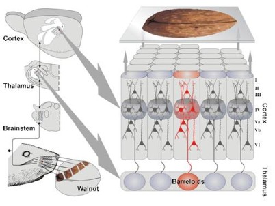
Electrophysiological and morphological investigations of neural networks in the rat primary somatosensory cortex (barrel-cortex).
The main research interest of our group is the investigation of structural and functional (re)organization of cortical networks in the rodent somatosensory (barrel) cortex in health and disease. The scaffold of proper structural and functional organization of the cortex – and thus eventually its accurate performance while receiving, processing and sending information - is depending on the activity of a multitude of different transcription factors, growth factors and neuromodulators during brain development.
Many neurological disorders and their related cortical dysfunctions can be linked with abnormal activity of one or more factors or neuromodulators - be it because of genetic variations or because of pharmacological modulation during critical periods of brain development. The links between brain development and cortical performance in terms of accurate or distorted receiving, processing and sending of information, can be studied very well in the distinctively organized sensory systems and the respective primary sensory cortical areas.
In our group, we focus on the structural and functional organization of the neural networks in the rodent primary somatosensory cortex.
On the one hand, we aim to improve our fundamental understanding of the somatosensory system.
On the other hand, we use this structurally and functionally well-organized sensory system as model for answering general questions about (i) how certain gene defects (e.g. mutations that are linked to autism spectrum disorders) or (ii) how altered levels of neuromodulators (e.g. serotonin) or neurotrophic factors (BDNF) can affect cortical network organization and how this can potentially explain related symptoms and behavior.
To gain insight into the structural and functional properties of the somatosensory cortical networks we use electrophysiological and neuroanatomical in vitro approaches on network and single cell level in mouse as well as rat models. The question which function the numerous diverse cortical neuron-classes have in processing sensory information, may only be answered by methods that reveal detailed information about (i) how single neurons are structurally and functionally integrated in complex cortical circuits and (ii) how populations of neurons interact with each other. To reveal the functional properties we combine (paired) patch clamp recording with optical stimulation techniques (caged glutamate photolysis and recently optogenetics) and multi-electrode array (MEA) recordings.
We investigate the structural aspect of cortical network organization by tracing studies and 3-dimensional single neuron reconstructions. The intense collaborations with local and international research groups from molecular neurobiology, behavioral neuroscience and neuroinformatics allow studies on an interdisciplinary level covering aspects from gene to behavior as well as modeling.
Current projects deal with investigating the impact of altered 5-HT levels on the development and organization of the somatosensory system, the effects of BDNF splice variant expression on synaptic plasticity, signal integration and oscillatory activity between neural networks, and developmental control of cortical parcellation and laminarization.

In acute coronal slices (background) visually identified neurons are electrophysiologically characterized. After that, inactive caged-glutamate is added to the bath solution. A UV flash is focused through the objective onto numerous different cortical fields of 50x50 µm in size. In these fields, very locally, caged glutamate is photolysed to active L-glutamate which activates local neurons.
With computer controlled methods a large cortical area (grid) is mapped without gaps for any origins of monosynaptic excitatory and inhibitory inputs. The spatial resolution is sufficient to reveal not only layer-, but also sublayer-specific differences in functional connectivity.
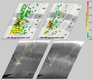
Photomicrographs of the native slices with positioned recording electrode (bottom) and, respectively, drawings of their columnar and laminar organization (top) are superimposed by the somatodendritic reconstructions of the recorded neurons and the maps of origins for synaptic inputs.
The maps illustrate the origins for excitatory (green to red) inhibitory synaptic inputs (blue) onto two morphologically and electrophysiologically heterogeneous pyramidal cells.